

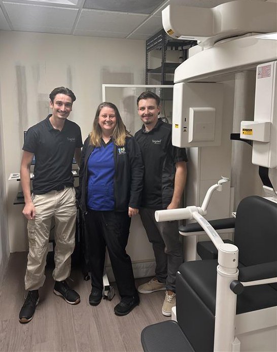
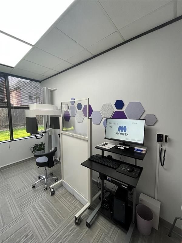
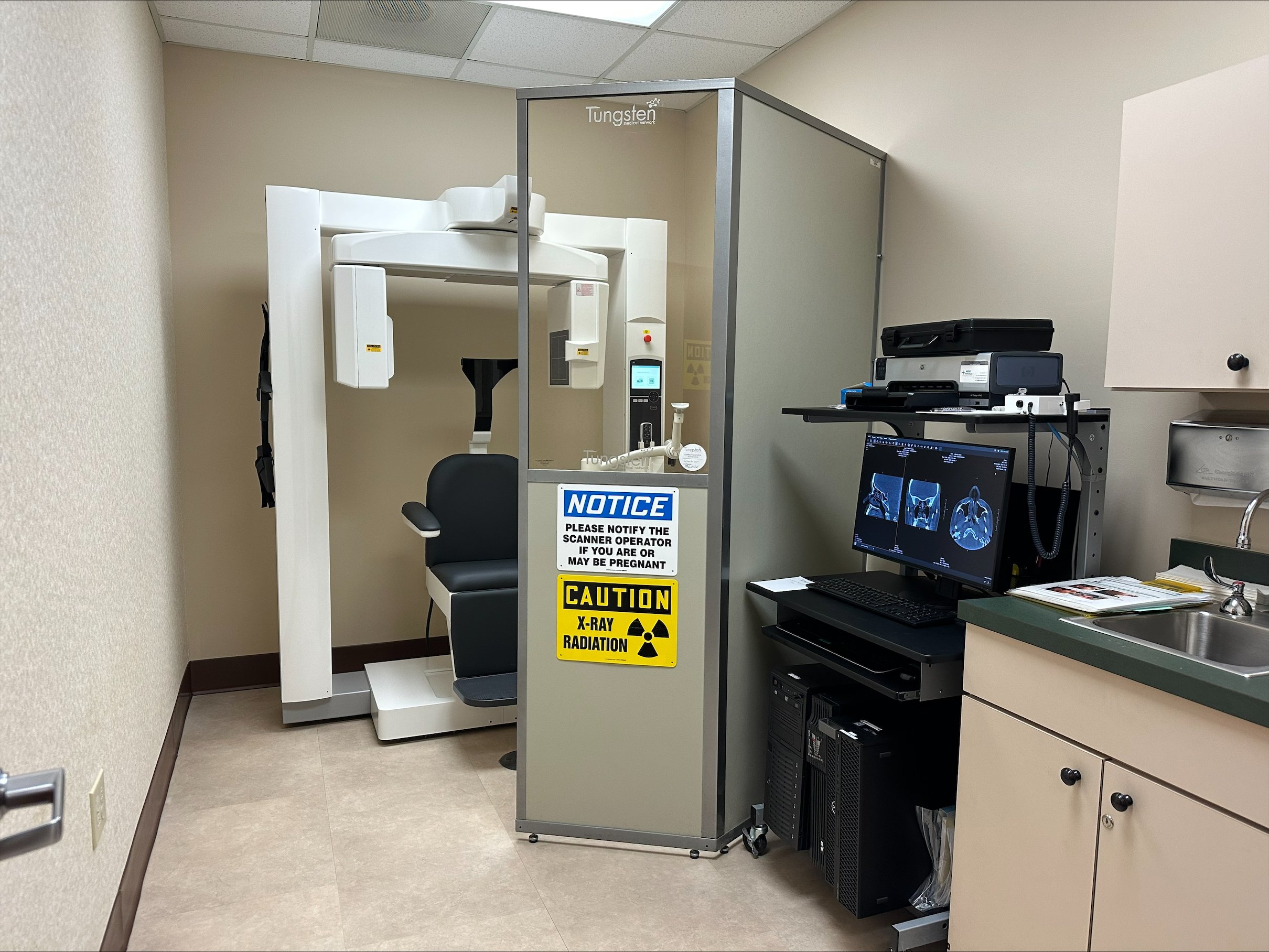
In-Office CT Solutions

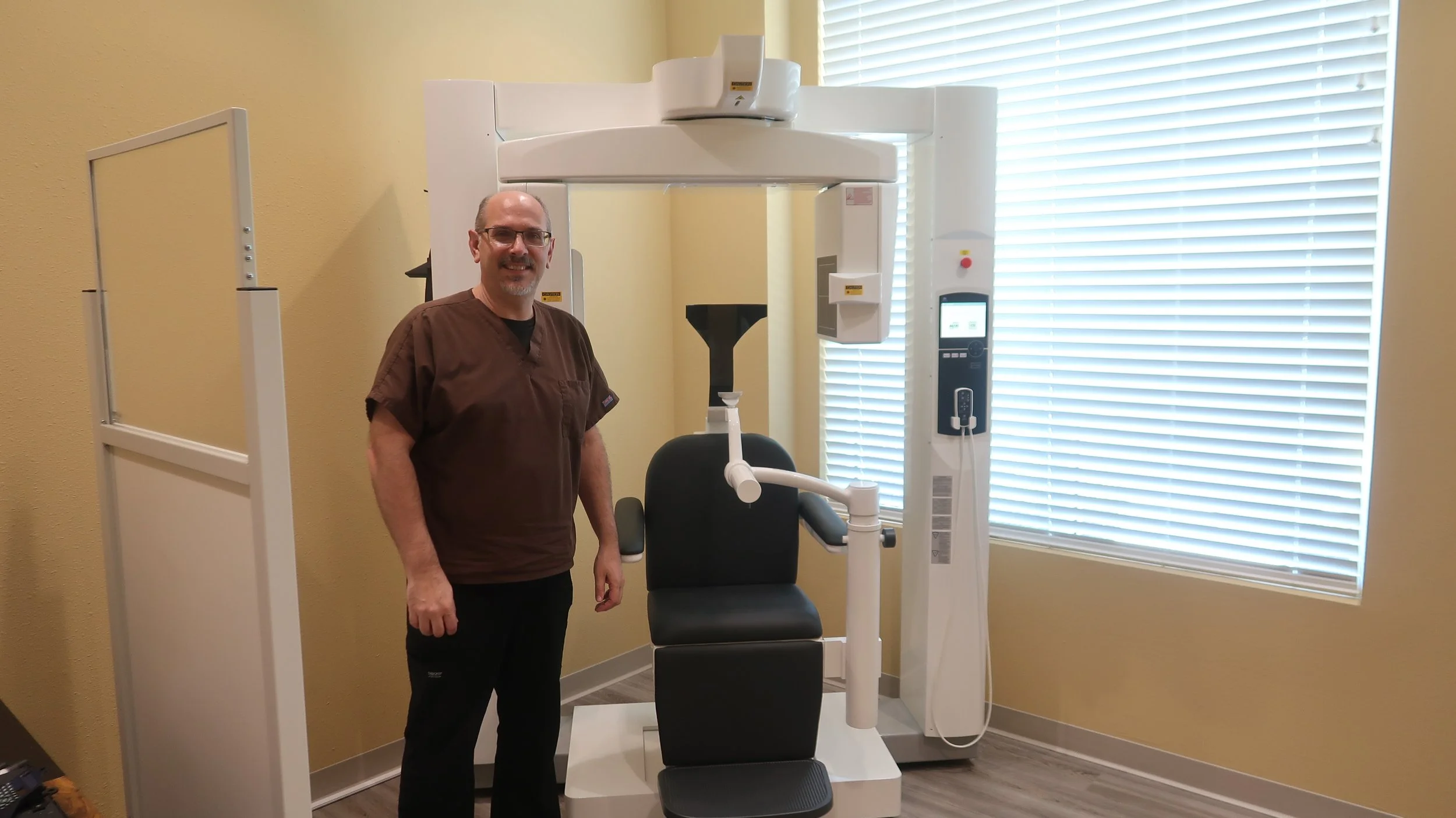
J. Morita 3D Accuitomo 170
With unparalleled quality, the Accuitomo offers unsurpassed image clarity and years of proven use in the market, making it a sustainable and sophisticated solution that can fulfill all your diagnostic needs effectively.

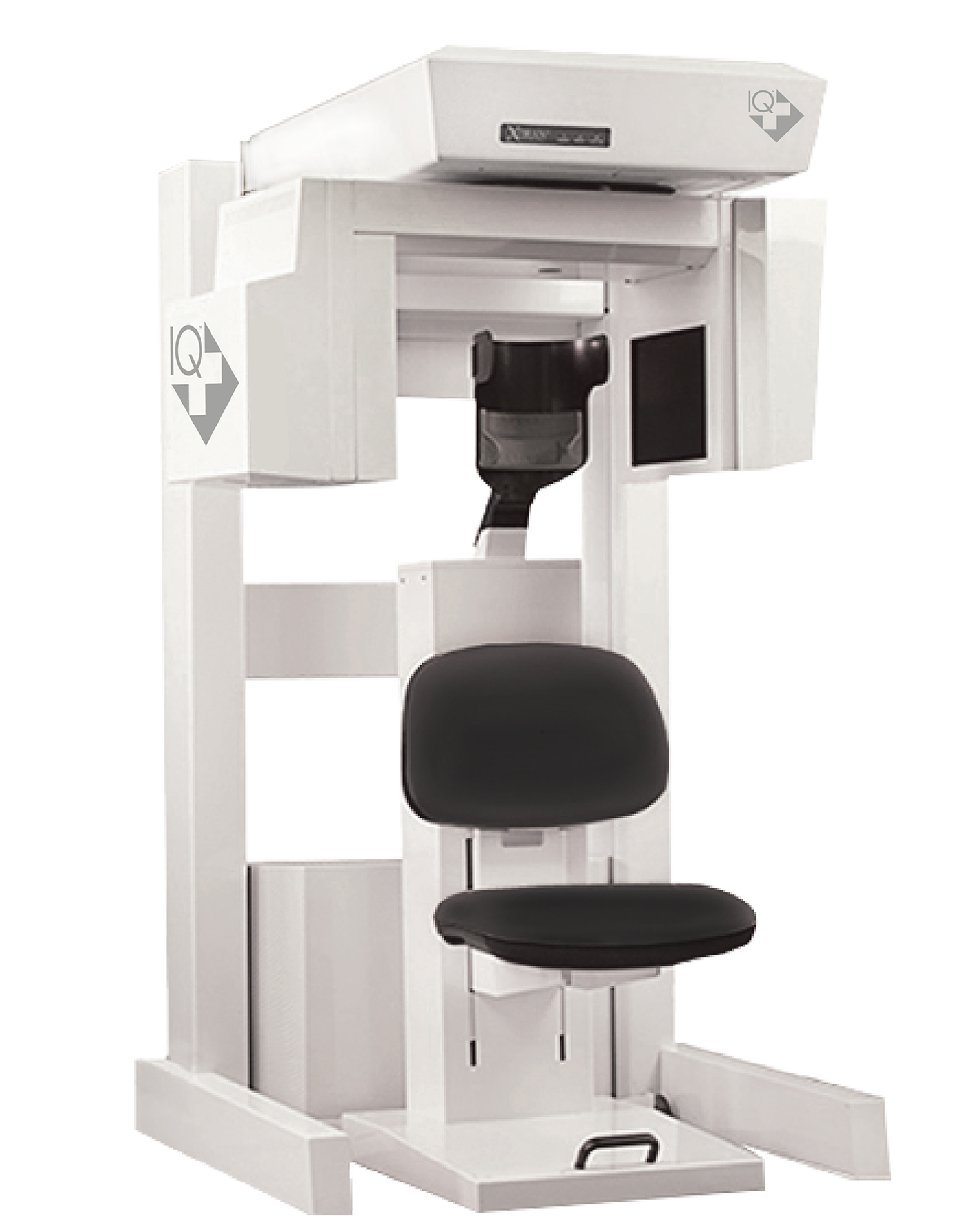
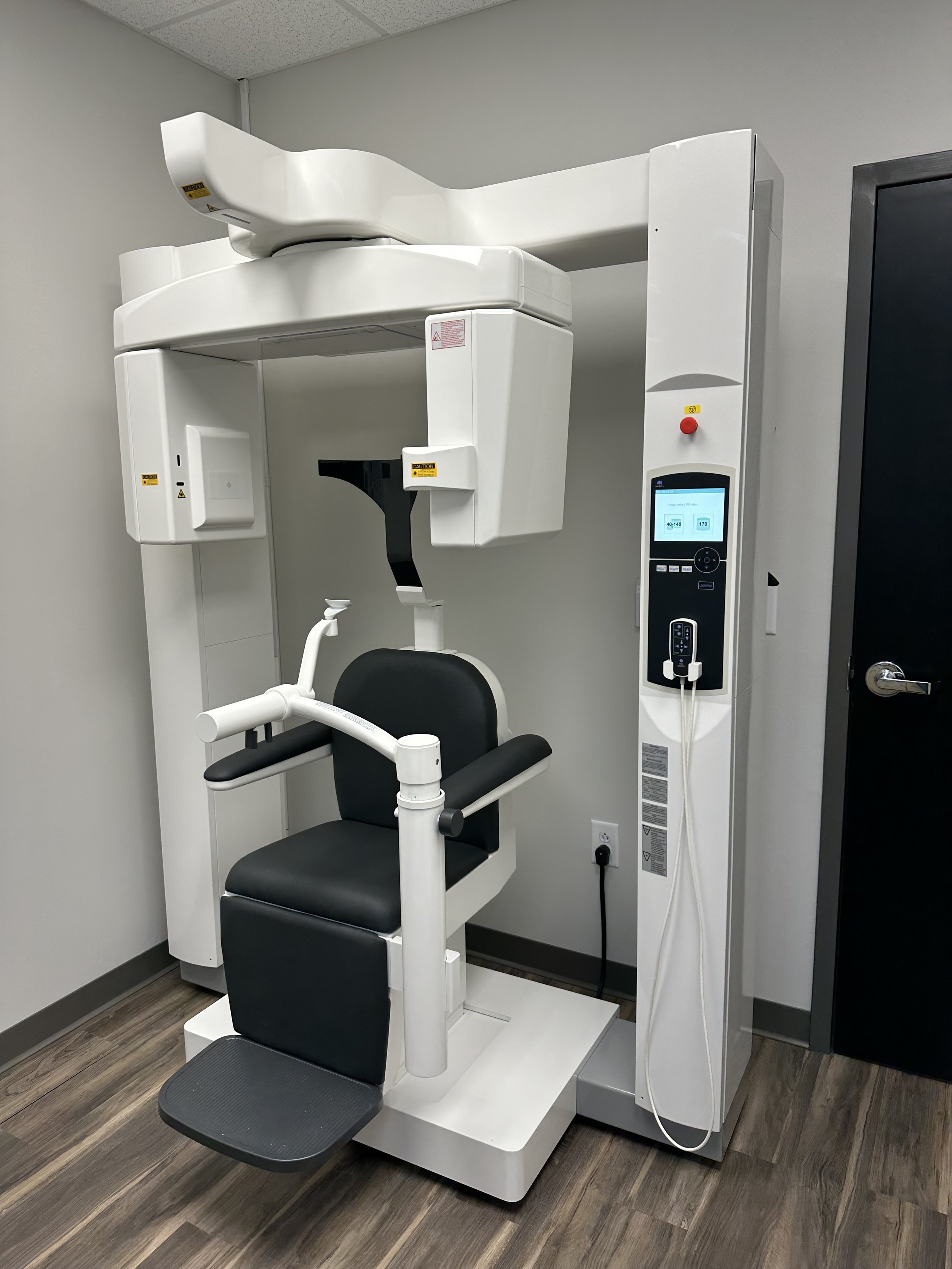
J. Morita FX800
The compact size, wheelchair accessibility, and adjustable control panel of the FX800 make it an ideal choice for limited spaces and ensuring patient versatility.
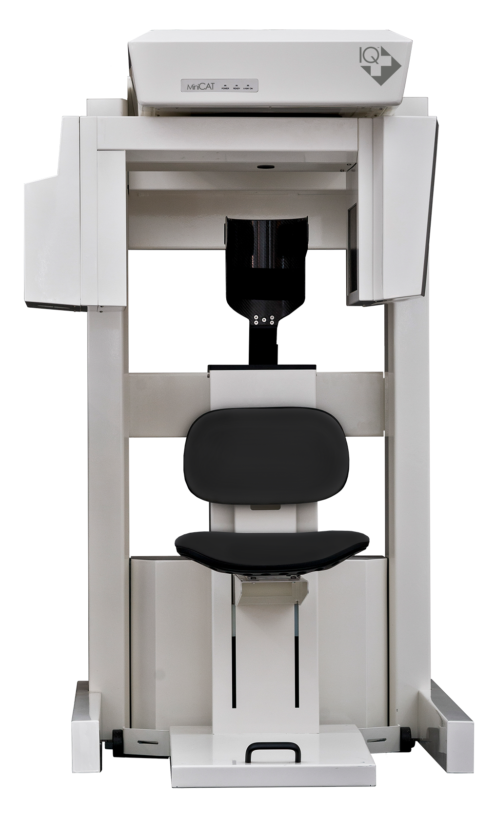
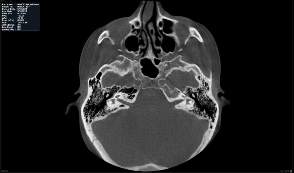
Xoran MiniCAT IQ+
The future of imaging with Next Generation MiniCAT IQ
Plus CT scanner:
Enhanced image quality
and lightning-fast scans for unparalleled diagnostics.
The Morita 3D Accuitomo CT Scanner
Morita’s Most Advanced CBCT Unit
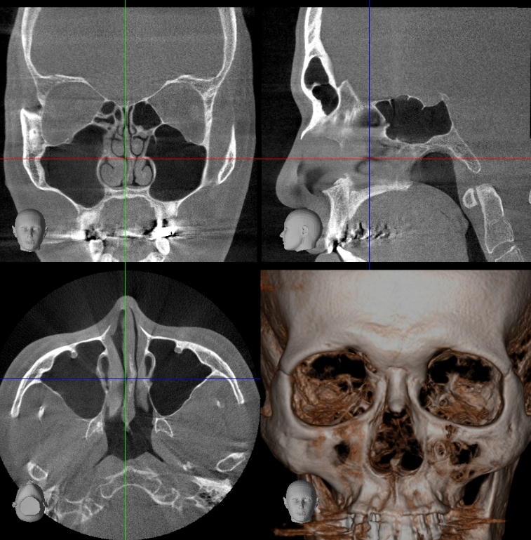
The Accuitomo CT scanner is Morita’s most advanced unit. Highly refined, it is the 4th generation of the Accuitomo product line. It offers a voxel size of just 80 µm and displays even the most subtle details of bone structure. This super-fine voxel combined with the unit’s 14 bit grayscale capability creates a wide dynamic range which produces the highest quality visualization of both hard and soft tissue areas. It enables a comprehensive examination for diagnosing sinus and temporal bone pathology.
-
With the flat panel detector, high-quality and detailed Images of the structures of the head and neck can be generated. Adjusting the position of the FPD reduces X-ray dose, provides higher resolution, and minimizes distortion.
Optimizing collimation of the beam, depending on the size of the area, also reduces X-ray dose and X-ray scattering.
-
There are nine sizes for exposure regions with diameters ranging from 40 mm to 170 mm.
Ø 170 x 120 mm
Ø 170 x 50 mm
Ø 140 x 100 mm
Ø 140 x 50 mm
Ø 100 x 100 mm
Ø 100 x 50 mm
Ø 80 x 80 mm
Ø 60 x 60 mm
Ø 40 x 40 mm
Resolution stays high and distortion is minimized for all regions from the smallest (ø 40 x 40 mm) to the largest (ø 170 x 120 mm).
-
High Resolution Mode (Hi-Res)
This is the highest resolution. Exposures are made at one-fourth the size of the detector pixels for the greatest spatial resolution. Ideal for observation of delicate bone structures such as the ossicular chain.High Fidelity Mode (Hi-Fi)
This mode has high data density data to make clearer and sharper images. This is especially good for performing zoom reconstructions.High Speed Mode (Hi-Speed)
Full scan: 10.5 sec. Half scan: 5.4 sec.Reduces motion artifacts. Good for children or others with difficulty remaining motionless.
Standard Mode (Std)
Suitable for limited and wide views of temporal bone, paranasal sinus, maxilla and mandible, individual teeth, etc. -
The scout positioning system is easy and accurate. Use the triple beam positioning system for even greater precision.
Two-Direction Scout
The region of interest can be easily targeted by making images from two directions. Then you can simply click on the images to specify the center of the region of interest. This information is transmitted to the X-ray unit, and the chair automatically moves into position.
Use scout to accurately determine the minimal region of interest before exposing the patient to the higher dosage CT scan.
Easy High Precision
The region of interest can be easily targeted using the three positioning laser beams. The patient’s head is safely and securely stabilized by the chinrest and headrest.
-
Installing i-Dixel software on all intra-clinic computers enables sharing of image data on each linked client computer. Observation of images on non-network computers can be achieved with the One Data Viewer, and the One Volume Viewer without installing i-Dixel.
One Data Viewer & One Volume Viewer Software
These unique Morita applications let you view three dimensional images and volume rendered images even if the computer does not have i-Dixel software installed.
CT data can be exported from the i-Dixel application and later stored on a DVD. This DVD can then be used on a computer outside the clinic to view CT images, volume rendered images and patient information.
Additional functions include zoom, black and white reverse, brightness, and contrast adjustment as well as optional length and angle measurement capabilities.
i-Dixel conforms to the following DICOM standards:
1.Modality worklist management service class (optional)
2. Storage service class
3. Modality performed procedure step service class
4. Print management service class
The FX800 is a CBCT X-ray unit that produces stunning images for ENT evaluation. It is intended for the examination and diagnosis of the skull including the ENT region. High resolution, this unit offers a minute voxel size of just 80 μm and features an adjustable horizontal X-ray beam for artifact reduction. Two exposure modes offer control and flexibility with a 360° high definition scan, or a faster 180° rotation with reduced dose. The FX800 has reached the pinnacle of CBCT imaging technology.
The Morita FX800 CT Scanner
The New Frontier for CBCT Imaging
-
For FOV Ø 40 × H 40 exposures, the voxel size is 80 μm and resolution is 2.5 LP/mm.
Spatial resolution indicates how small objects can be and still been discriminated visually. This is called spatial frequency and is usually expressed as “line pairs per millimeter (LP/mm)”. This indicates how many pairs of white and black lines can be discriminated within 1 millimeter; the higher the number, the greater the resolution. MTF (Modulation Transfer Function) is one way to objectively evaluate the line-pair resolution and objectively express how many line-pairs and at what level of contrast can be discriminated. Generally, if MTF is 10%, naked eye discrimination is possible. Spatial resolution does not depend only on voxel size.
-
For the first time, Morita’s Zoom Reconstruction feature is available on a multi-functional unit. After taking an image with a voxel size of 125 μm, reconstruction can be repeated for a higher resolution of 80 μm voxel size without retaking the exposure.
-
A new taller field of view makes DICOM registration with surgical navigation systems even easier and more accurate.
-
Face-to-Face Positioning
Laser beam positioning is more accurate if you have good communication with the patient.
-
Control Panel
The control panel moves freely so that it can be used from the front or side, for improved access during patient positioning.
-
Wheelchair Compatible
The chinrest can be lowered to approximately 34″ (865 mm) (short column) to accommodate patients in wheelchairs.


Xoran MiniCAT IQ+
The Next Generation MiniCAT
Xoran’s flagship product, MiniCAT, is an in-office, point-of-care imaging solution designed specifically for ENT and Allergy physicians. ENTs rely on MiniCAT because it offers low-dose, contact-free imaging for patients, supports same-day diagnoses, and enhances treatment compliance—ultimately helping practices retain procedure revenue.
-
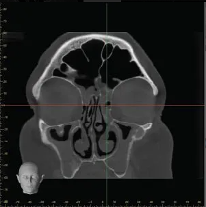
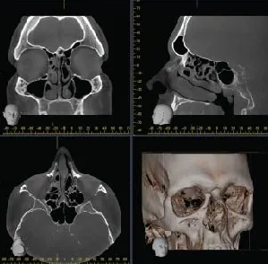
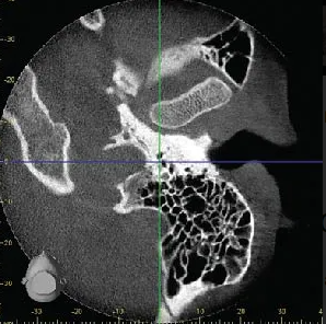
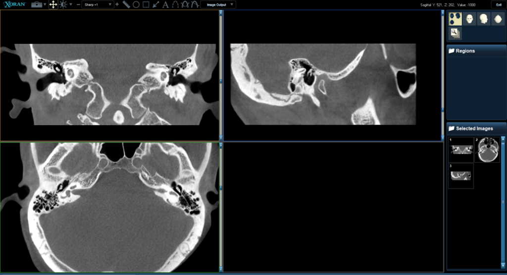
0.07 mm Precision Imaging
Zoom in and rapidly reconstruct your region of interest (ROI) at 70 microns.
For areas like the sinuses, inner ear structures, and other complex anatomical regions, high-resolution zoom provides ENT specialists with the detailed imaging needed for more accurate diagnosis and effective treatment.
-
Xoran’s advanced ACE Mode assesses patient radiodensity, incorporates age-specific considerations, and automatically generates a recommended imaging protocol—Small, Medium, or Large—tailored to each patient.
-
Physicians and healthcare providers have the final say in selecting the appropriate protocol for each patient. They can either follow the protocol recommended by ACE Mode or choose to override it, selecting a protocol based on the patient's specific clinical needs.
-
Doctors have full control over the timing and quality of their scans, ensuring patients receive both their diagnosis and treatment plan in a single visit.
The system integrates seamlessly with EHR and navigation systems, allowing scans to be correlated with billing records and streamlining the in-office diagnosis and treatment process.


What people are saying.